 |
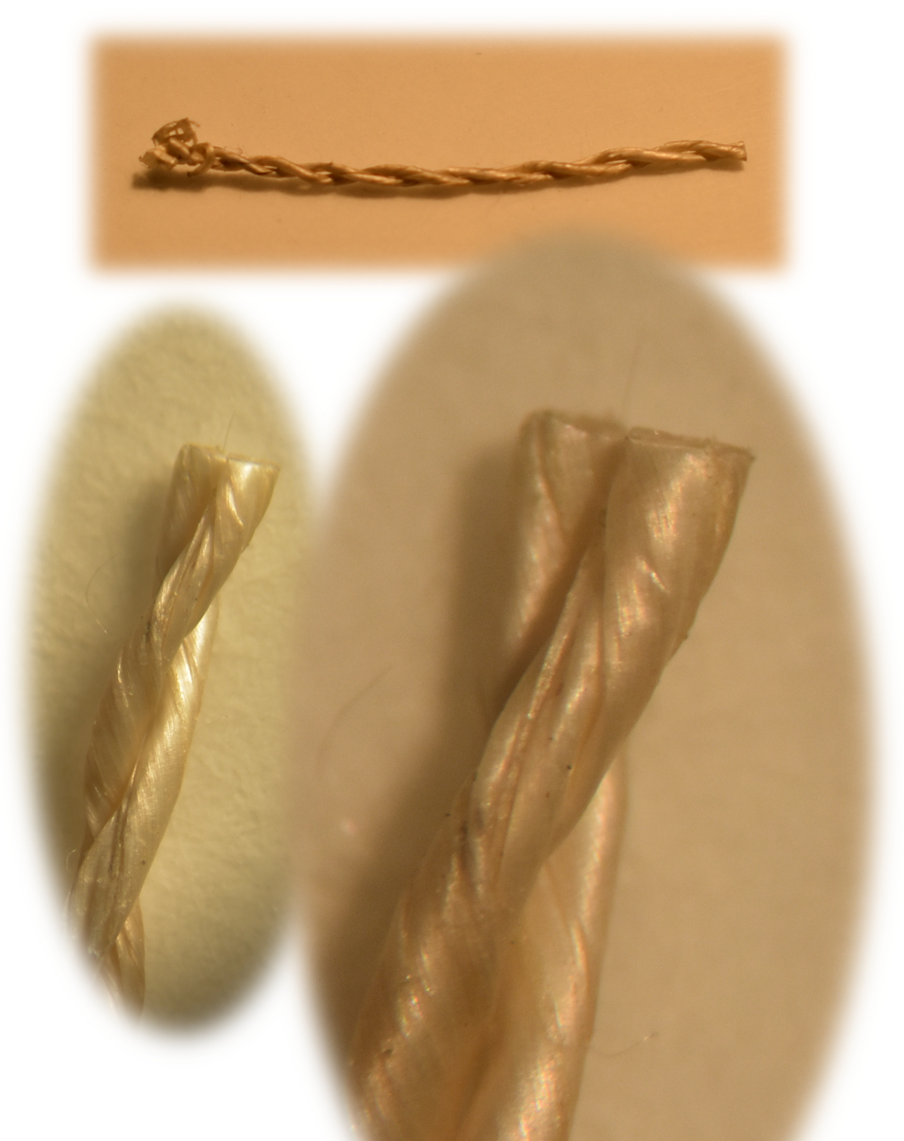
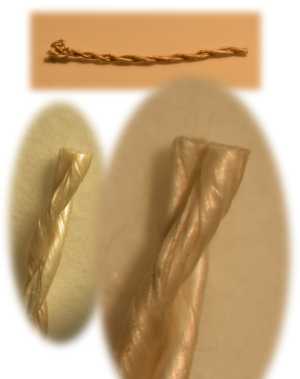
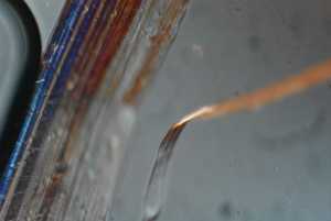
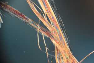
This composite image shows a single twisted or spun segment of yarn. You can see the two small single strands of yarn have been twisted around each other (i.e. two-ply) to make a relatively uniform and strong material. Original magnifications 1x, 10x, and 20x with incident incandescent light. |
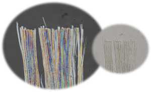 |
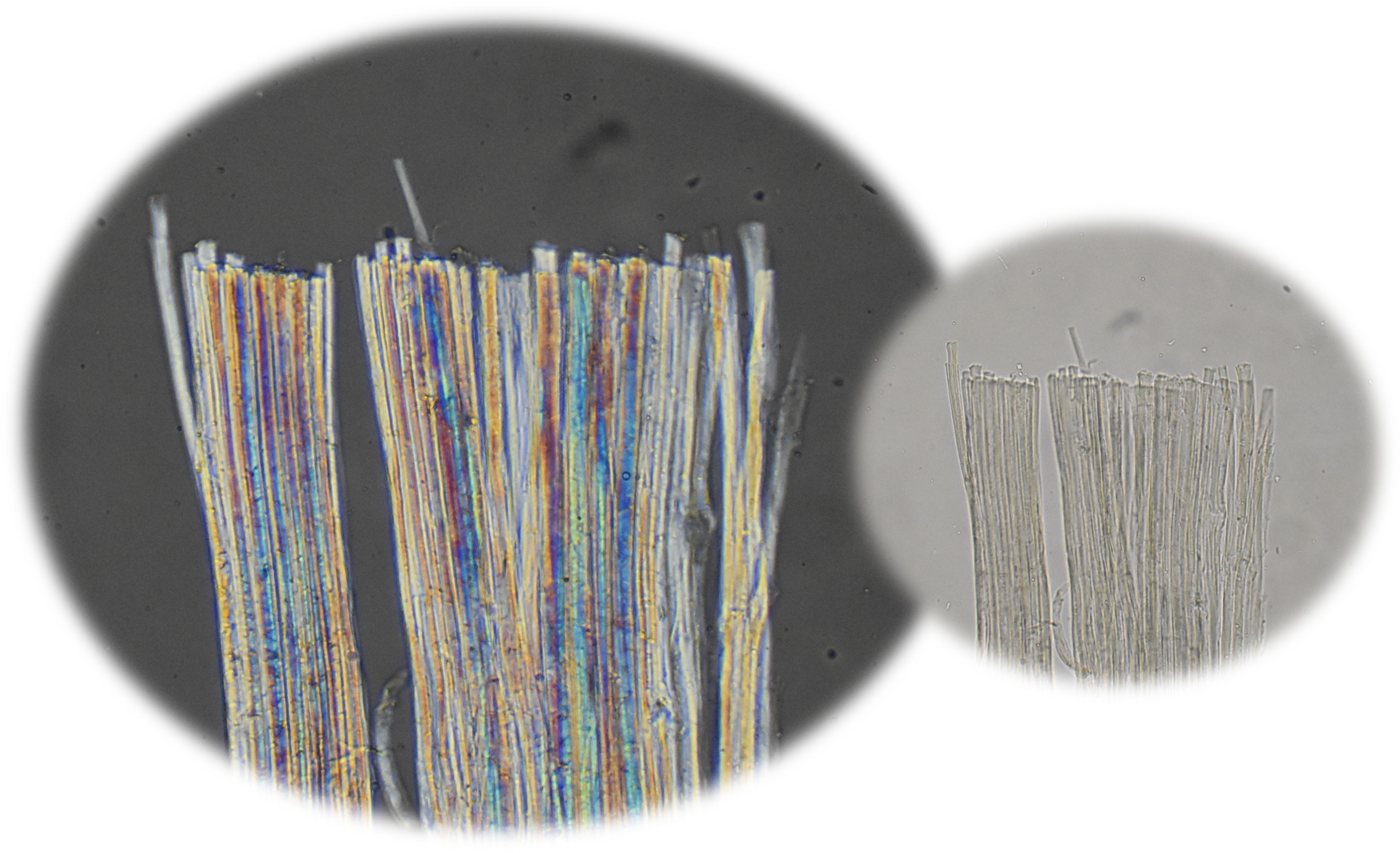
This composite image shows a bundle of Pueraria phaseoloides viewed with polarization microscopy (left panel) and routine transmission microscopy (right panel). Note the relative sharp margin of the fibers on the end where the bundle was cut with scissors. Polarization microscopy is useful in identifying internal structure...the various colors are due to the optical activity of the fibers and not intrinsic chemical differences in them. | |
 |
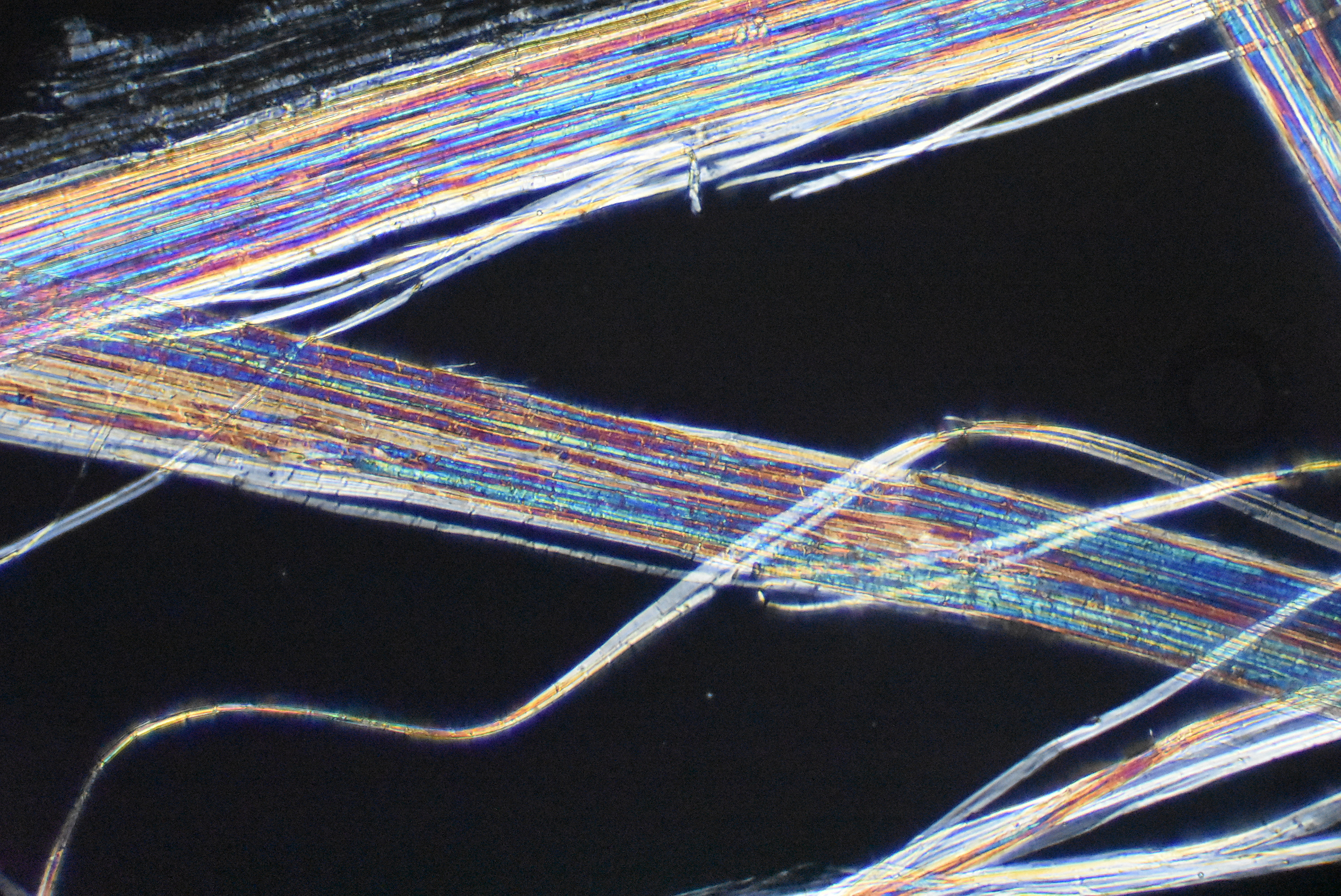
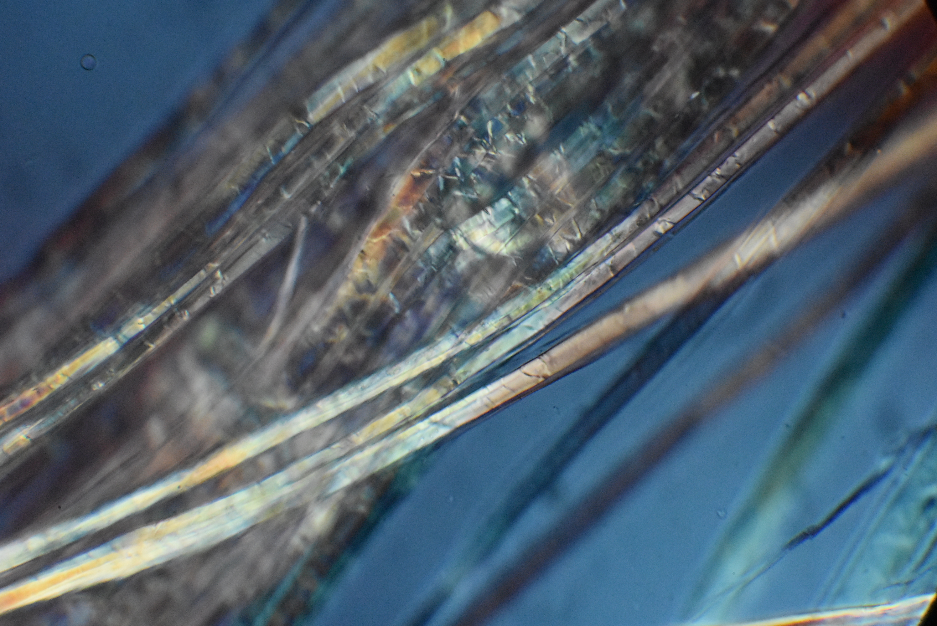
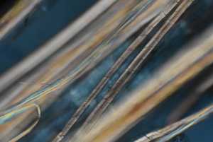
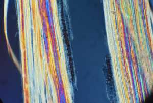
This is a high magnification view showing two larger bundles of fibers along with several individual ones. Notice how the edges of the bundles are relatively sharp and how the diameters of the individual fibers change as they slightly twist or change direction. The change in apparent diameter as the fibers change direction suggests the fibers are somewhat flat as opposed to round. |
 |
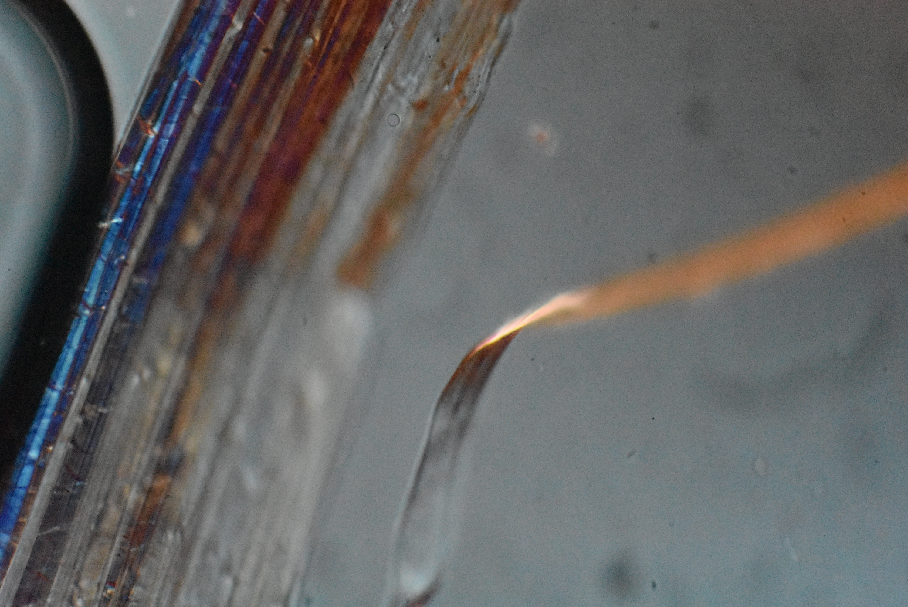
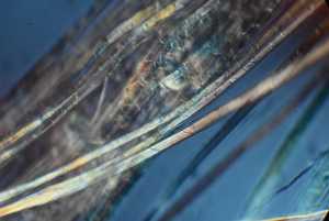
Notice the individual fiber on the right...it has a flattened appearance as it courses up and to the right; again suggesting a relatively flat fiber. Also notice the short cross hatches in the blue colored fibers on the left. Those may represent some surface kinking or other irregularities. The sharply circumscribed black object on the left is an air bubble. |
 |
Another high power magnification view showing bundle of fibers with some separating from the main bundle on the right side. Polarization microscopy view. |
 |
This is another polarization microscopic view showing several individual fibers exhibiting cross hatches. The outside of the fibers appears smooth. |
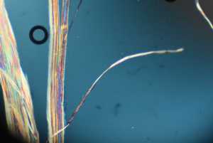 |
Another polarization microscopic view showing a single fiber in the foreground twisting slightly as it courses up and to the right. Again, the fiber appears somewhat flat. There is small air bubble at the upper left. |
 |
Polarization microscopic view showing several small bundles and two or three individual fibers. |
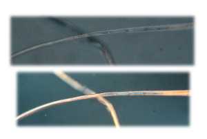 |
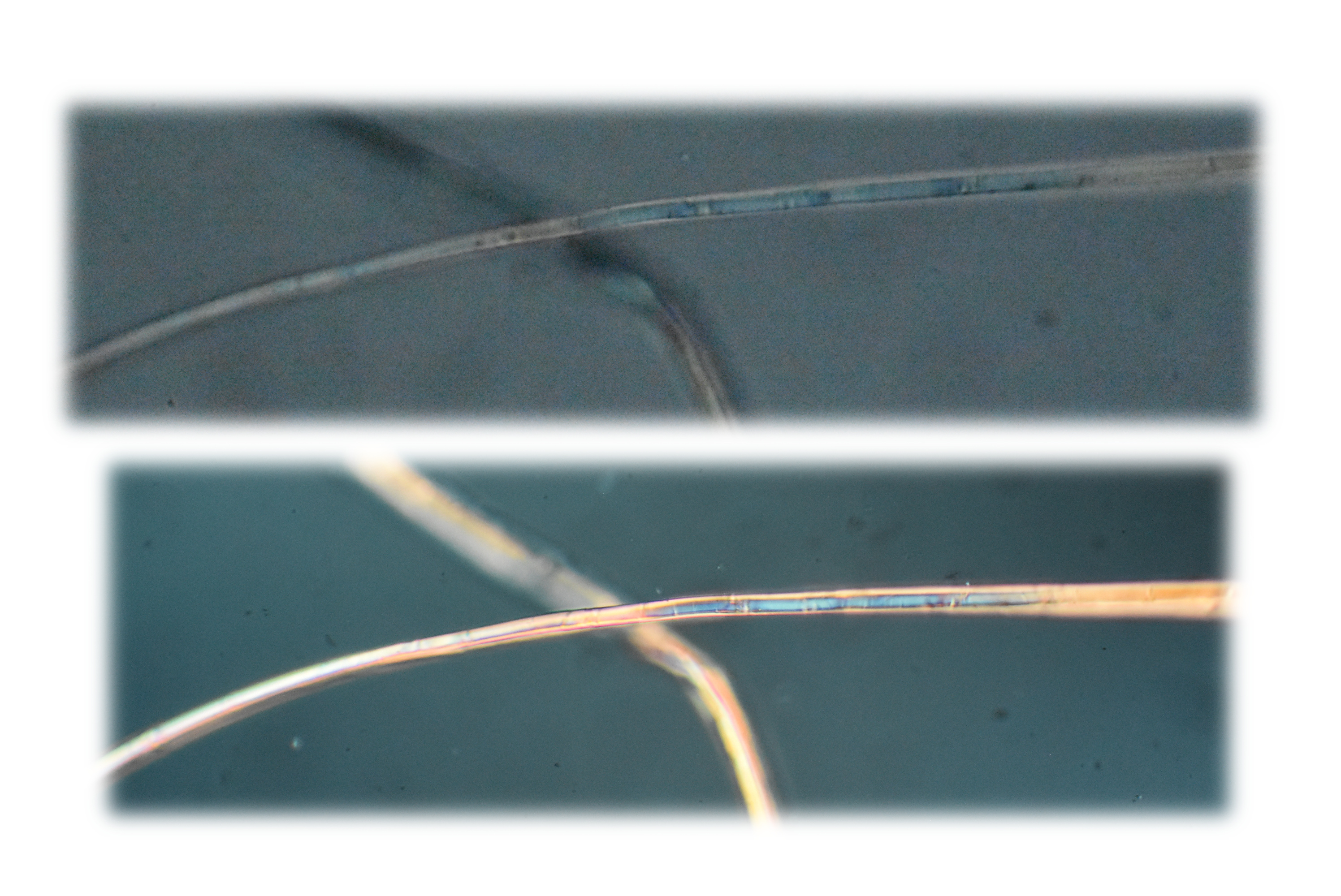
This composite image shows fibers using both conventional light microscopy (upper panel) and cross polarizing lenses (lower panel). |
 |
This image shows two larger bundles of fiber with some amorphous material that appears "stuck" on the outside. That may represent some debris, other contamination or might be part of the surface plant structure. |